Blog Archive
-
▼
2011
(48)
-
▼
March
(48)
- explosions in the sky the rescue mediafire
- picture of appendix location
- gitoformate
- prednisone tablets
- celebrities breastfeeding in public
- rubella syndrome
- iron fist
- chiropractic symbol
- documentation icon
- piperacetazine
- mn twins logo
- thioxanthines
- premature infant growth chart
- greater sulphur crested cockatoo
- hypophosphatasia
- staphylococcus bacterial infection
- spine diagram
- grass tetany
- fertilization cycle
- psychiatry logo
- spain beach women
- lung cancer treatment
- oxygen tank and mask
- posterior pituitary hormones
- minor frostbite face
- bottom inspectors
- magnetic permeability table
- alcohol units
- trachea esophagus diagram
- job description for cashier
- arabian peninsula physical map
- murdered out cars for sale
- autonomic nervous system receptors
- pictures of chlamydia in the mouth
- phenformin
- pokemon genosect
- seaweed salad calories
- examples of expert systems
- juvenile arthritis symptoms
- personality development logo
- perindopril
- oral hygiene for children
- science hazard warning signs
- delirium sandman
- telangiectasia face
- econazole nitrate
- clean rooms international
- insulin lispro
-
▼
March
(48)
Wednesday, March 9, 2011
trachea esophagus diagram

Thumbnails: Medical diagrams and resources regarding Digestive System.

passages (trachea (windpipe) and lungs) and the esophagus (eating tube).

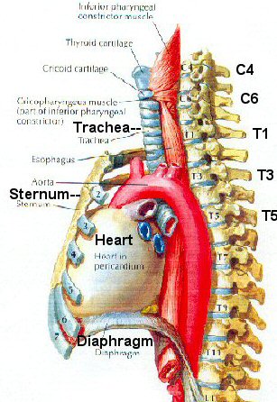
of structures such as the trachea, esophagus, heart, and major vessels,

The trachea is rigid, while the esophagus is quite soft and flexible,

An illustration of the isolated, innervated guinea pig trachea/esophagus

Below: Larynx, Trachea, Esophagus

Match the number in the diagram with the part listed below:

Oh okay I'll put the diagram here, but there is more info on that previous

is last vessel from arch and extends dorsal to trachea and esophagus.

Main article: Tracheal tube

anatomy of the aorta in its relationship to the trachea and esophagus.

This diagram shows the aorta coursing to the right of the esophagus and

Continue separating the tissue with a probe until the trachea and esophagus

Rich had his food pipe (Esophagus) removed from the windpipe (Trachea) and

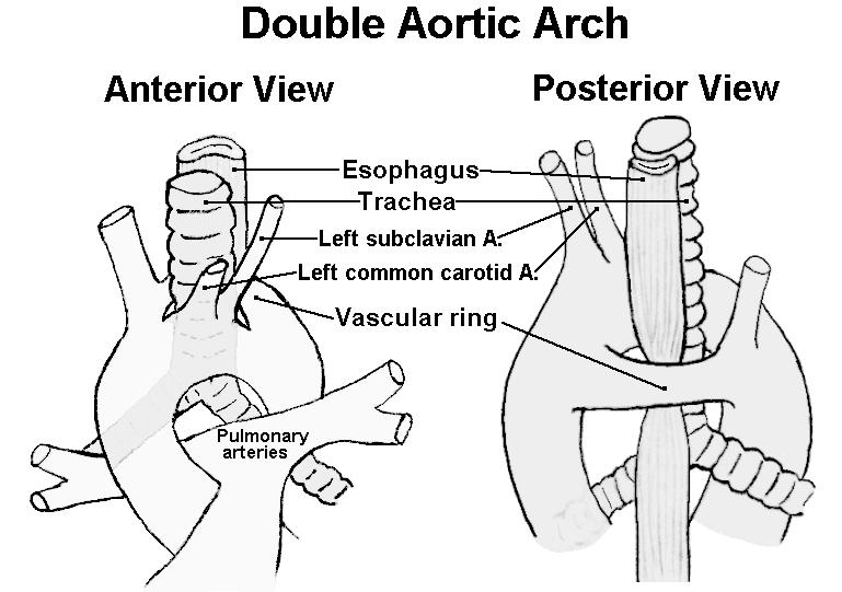
the two arches encircle the trachea and esophagus.

Nose; Pharynx; Larynx; Trachea; Bronchi; Lungs. Lets us now see the diagram

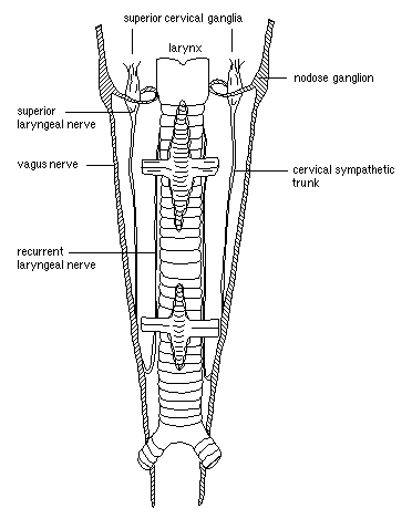
trachea; esophagus; left recurrent laryngeal nerve

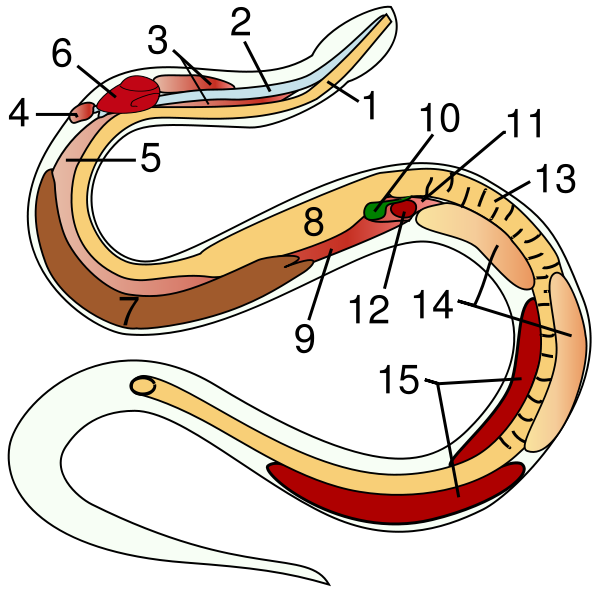
1 esophagus, 2 trachea, 3 tracheal lungs, 4 rudimentary left lung,

Section of skull, pharynx, epiglottis and larynx diagram

trachea, esophagus
Subscribe to:
Post Comments (Atom)
Followers
Powered by Blogger.
0 comments:
Post a Comment